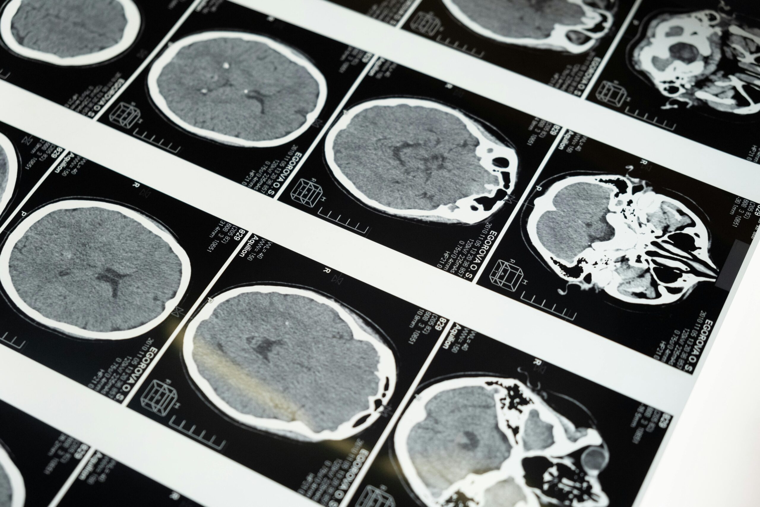
In the field of Artificial Intelligence, we often encounter the “Precision Gap.” In consumer tech, an AI might misidentify a cat as a dog with little consequence. In healthcare, missing the boundary of a tumor by even a few millimeters can change a patient’s surgical outcome. While general Deep Learning models are built to categorize images, U-Net was engineered for a far more difficult task: Semantic Segmentation. ## The Anatomy of Precision: The U-Net Architecture
The U-Net architecture, first introduced for biomedical data, operates on a unique “Encoder-Decoder” structure. Its name is derived from its symmetric U-shaped design, which balances two competing needs in medical imaging: Context and Location.
1. The Downsampling Path (The “What”)
The left side of the “U” acts as an encoder. It captures the global context of the image—identifying that it is looking at a lung, a brain, or a cell. However, as the model “shrinks” the image to understand its features, it loses the fine-grained spatial details.
2. The Upsampling Path (The “Where”)
The right side of the “U” acts as a decoder. It projects these abstract features back into the original dimensions of the image. Its goal is to create a pixel-by-pixel mask that matches the input perfectly.
3. The Power of Skip Connections
The true innovation of U-Net lies in its Skip Connections. These bridges pass high-resolution spatial information from the encoder directly to the decoder.
- The Result: The model doesn’t have to “remember” the exact edges of an organ; it can see them through these connections, allowing for ultra-sharp segmentation boundaries that other models simply cannot match.
Why It Outperforms in a Clinical Setting
Data Efficiency
Medical datasets are notoriously small because high-quality labels require hours of a specialist’s time. U-Net is remarkably robust; it uses heavy data augmentation (like elastic deformations) to learn effectively from limited samples. This makes it viable for rare diseases where only a few dozen scans might exist.
Pixel-Wise Accuracy
Unlike standard CNNs that provide a single label for an entire image, U-Net classifies every single pixel. For a radiologist, this means the difference between being told “there is a lesion” and having the lesion automatically outlined and its volume calculated instantly.
Adaptability: From 2D to 3D
The architecture is incredibly flexible. Whether it is a 2D X-ray or a complex 3D MRI volume (3D U-Net), the logic remains the same. Modern iterations, such as Res-UNet, now incorporate “Residual Blocks” from ResNet to allow for even deeper training without the risk of model degradation.
The Impact on Patient Care
U-Net has transitioned from a research paper to the backbone of modern surgical robotics, radiation therapy planning, and automated diagnostics. By bridging the gap between “seeing” an image and “understanding” its exact geometry, it has become an indispensable tool in the physician’s digital toolkit.
In an era where AI is often a “black box,” U-Net provides the transparency of a clear, visual map—exactly what clinicians need to make informed decisions.
#HealthcareAI #MedicalImaging #DeepLearning #Biomedical #MachineLearning #Radiology #UNet